


TECHNOLOGY & DEVELOPMENT
More than just watts
How the THERA-Trainer tigo Makes Joint Loads Visible During Cyclical Movement Training

Jakob Tiebel
Health Business Consultant
Training duration, cadence and power output are among the key parameters of cyclical movement training. However, they only offer limited insight into the actual mechanical loading occurring at the hip, knee and ankle joints. A study published in the Journal of Biomechanics therefore investigated how joint contact forces during seated movement training can be estimated using a model-based approach. In the experimental setup, the THERA-Trainer tigo served as a controllable, robotically driven movement therapy device.
The internal mechanical dose
Joint contact forces describe the resultant load within a joint. They are determined not solely by the force transmitted to the crank, but also by muscle activation, co-contraction, joint position, force application, anatomical lever ratios and movement coordination. This means that two individuals can generate markedly different internal loads despite identical power output and cadence. In particular, radial crank forces and muscle forces can place strain on the joints without this being directly reflected in the displayed wattage.
Since joint contact forces can only be measured directly using invasive procedures, the researchers combined experimental measurement data with an individualised neuromusculoskeletal computer model.
Since joint contact forces can only be measured directly using invasive procedures, the researchers combined experimental measurement data with an individualised neuromusculoskeletal computer model.
The Experimental Setup with the THERA-Trainer tigo
Eleven healthy adults took part in the investigation. Participants trained on a THERA-Trainer tigo under six controlled conditions:
• 40 or 60 revolutions per minute
• 15, 30 or 45 watts each
Position, seat spacing and movement execution were standardised. Specialised cranks recorded tangential and radial forces as well as crank angle. In addition, movements were recorded using a three-dimensional camera system, and the activity of 15 muscles was captured using surface EMG.
The measurement data were incorporated into a computer model adapted to individual body height and body mass. On this basis, muscle forces as well as the contact forces at the hip, knee and ankle joints were estimated.
The THERA-Trainer tigo did not itself measure the joint forces during this process. It provided the reproducible movement task and standardised human-machine interface necessary for biomechanical analysis.
• 40 or 60 revolutions per minute
• 15, 30 or 45 watts each
Position, seat spacing and movement execution were standardised. Specialised cranks recorded tangential and radial forces as well as crank angle. In addition, movements were recorded using a three-dimensional camera system, and the activity of 15 muscles was captured using surface EMG.
The measurement data were incorporated into a computer model adapted to individual body height and body mass. On this basis, muscle forces as well as the contact forces at the hip, knee and ankle joints were estimated.
The THERA-Trainer tigo did not itself measure the joint forces during this process. It provided the reproducible movement task and standardised human-machine interface necessary for biomechanical analysis.
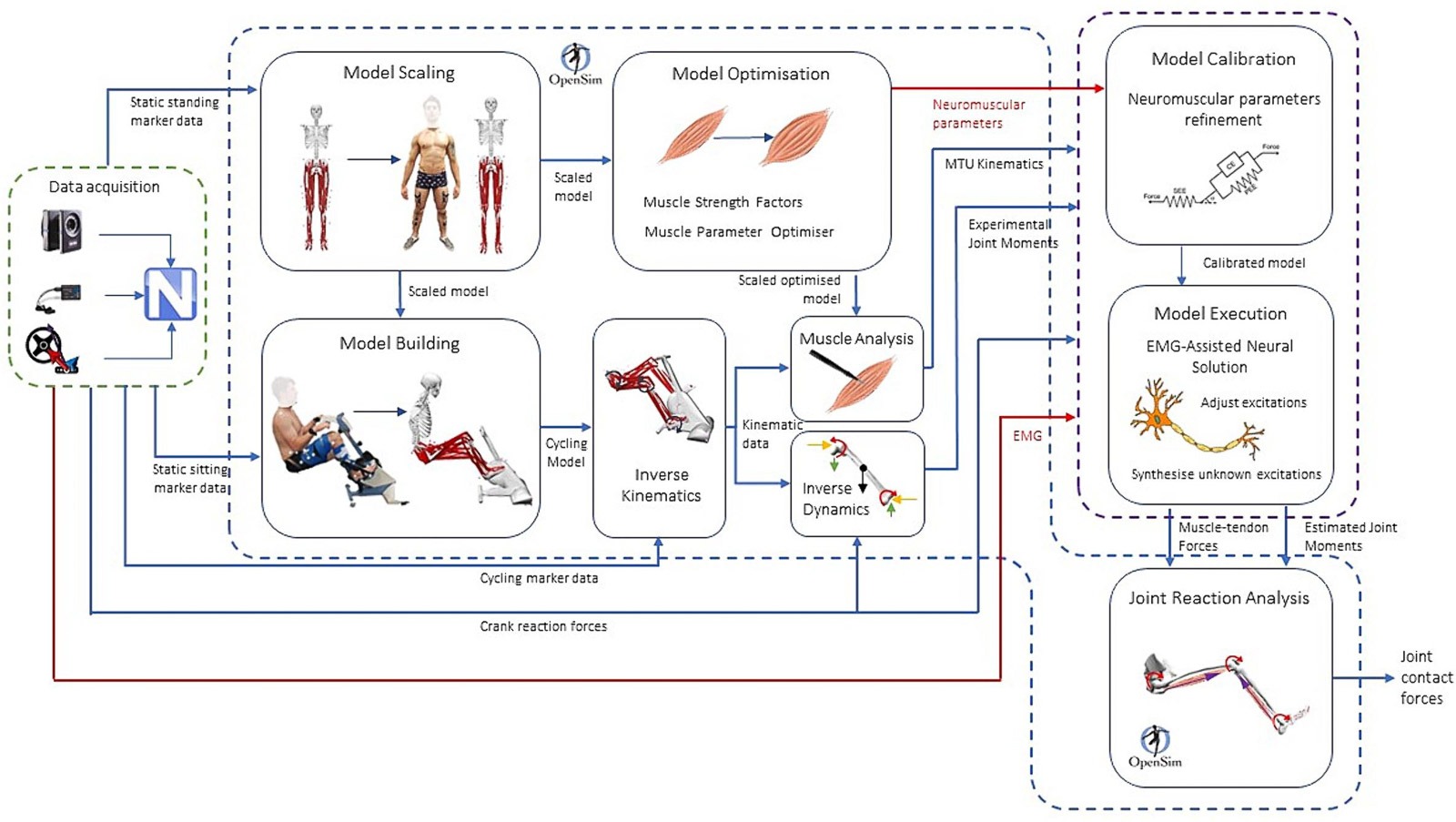
The diagram illustrates the data-processing workflow, from measurement through to biomechanical analysis. Time-synchronised muscle activity (EMG), movement data and crank forces were processed and subsequently incorporated into a subject-specific musculoskeletal model. From this, muscle forces and the resulting joint contact forces at the hip, knee and ankle joints were calculated using the modelling approach.
Same power output, different joint loading
The key finding: at identical power output, peak contact forces at group level were higher at 40 rpm than at 60 rpm. This effect was observed at the hip, knee and ankle joints. The difference was most pronounced at the knee. At the knee, mean peak contact forces at the lower cadence were approximately 0.33 to 0.46 body weights higher, depending on the power level.
This is biomechanically plausible: if the same power output is to be achieved at a lower cadence, a higher torque per revolution is required. The resulting joint loading, however, also depends on which muscles are activated, the extent to which antagonistic muscle groups co-contract, and the timing and direction of the force applied to the crank.
Accordingly, mean crank power output showed only a weak association with peak joint contact forces. The correlation values ranged between 0.063 and 0.211. A reliable estimation of internal joint loading based on power output (watts) alone was therefore not possible.
This is biomechanically plausible: if the same power output is to be achieved at a lower cadence, a higher torque per revolution is required. The resulting joint loading, however, also depends on which muscles are activated, the extent to which antagonistic muscle groups co-contract, and the timing and direction of the force applied to the crank.
Accordingly, mean crank power output showed only a weak association with peak joint contact forces. The correlation values ranged between 0.063 and 0.211. A reliable estimation of internal joint loading based on power output (watts) alone was therefore not possible.
Individual mobility strategies remain crucial
The group means did not hold true for every individual. In some participants, higher loads at the knee or ankle joint occurred at 60 rpm. The timing of peak joint loading within the crank cycle also varied.
This variability points to individual neuromuscular strategies. Individuals can accomplish the same external movement task through differing muscle recruitment, force distribution, stabilisation and co-contraction.
Consequently, no generalised dosage rule can be derived from these findings. A higher cadence is not inherently safer, nor is a lower cadence inherently more strenuous. The individual biomechanical response to the specific training condition is what matters.
This variability points to individual neuromuscular strategies. Individuals can accomplish the same external movement task through differing muscle recruitment, force distribution, stabilisation and co-contraction.
Consequently, no generalised dosage rule can be derived from these findings. A higher cadence is not inherently safer, nor is a lower cadence inherently more strenuous. The individual biomechanical response to the specific training condition is what matters.
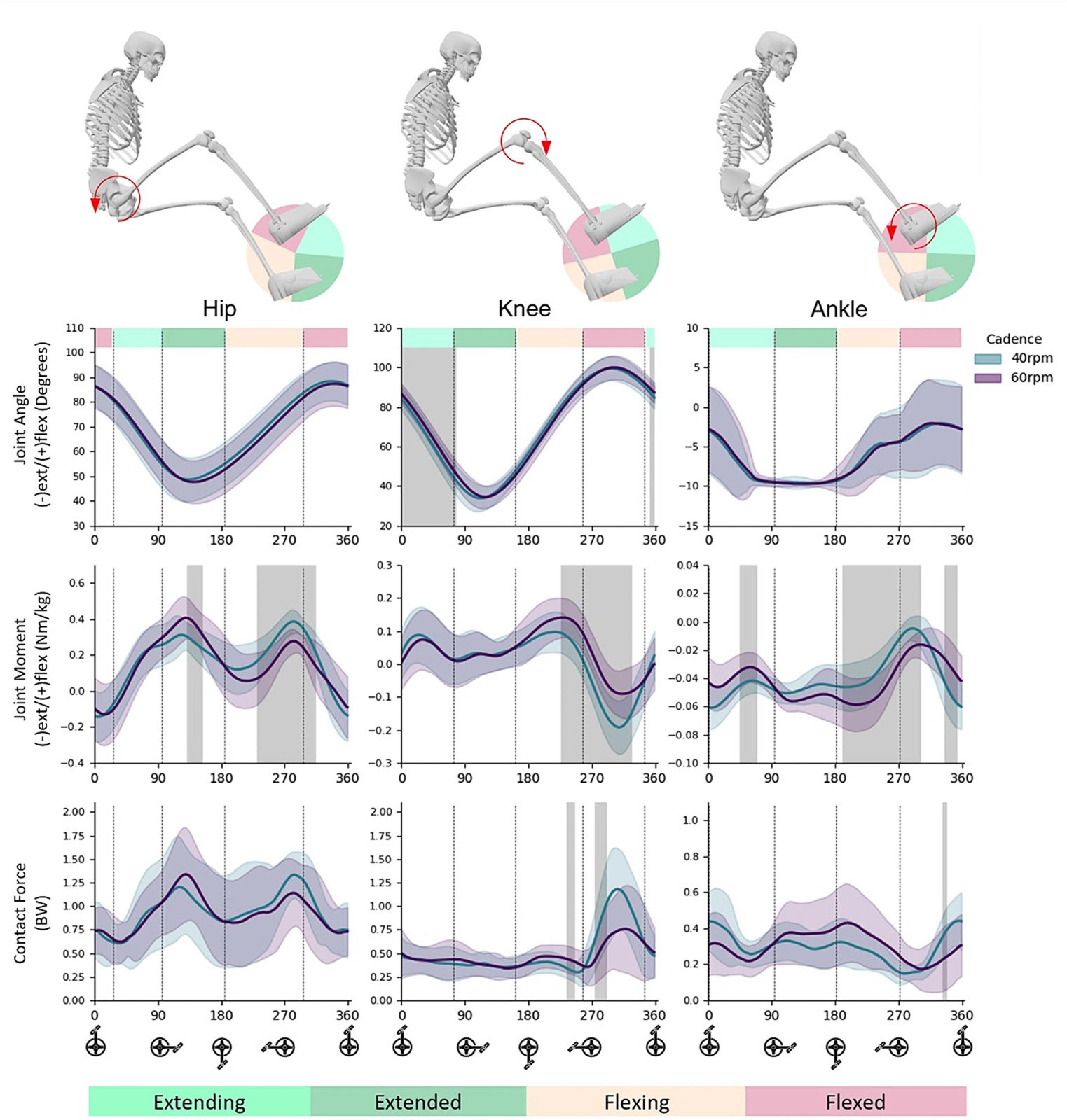
The diagram illustrates movement trajectories (top), joint moments (middle) and joint contact forces (bottom) at the hip, knee and ankle joints during cycling at 40 and 60 rpm, at comparable power output (~15 W). The values are shown over one complete crank revolution. Grey-shaded areas indicate significant differences between the two cadences (p < 0.05).
Prospects for research and training management
The study broadens the perspective on training dosage. Common parameters such as duration, resistance, cadence, power output and movement cycles remain important, but do not fully capture internal mechanical loading.
In conjunction with additional sensors and patient-specific models, it may become possible in future to estimate joint loading during training. For FES-assisted applications, it is also conceivable that stimulation patterns could, in the longer term, be tailored to the desired mechanical loading.
This is a promising direction for research and development. Real-time estimation of joint forces or automated model-based control of functional electrical stimulation was neither clinically validated nor investigated as a standard feature of the THERA-Trainer tigo within the study.
GET IN TOUCH
In conjunction with additional sensors and patient-specific models, it may become possible in future to estimate joint loading during training. For FES-assisted applications, it is also conceivable that stimulation patterns could, in the longer term, be tailored to the desired mechanical loading.
This is a promising direction for research and development. Real-time estimation of joint forces or automated model-based control of functional electrical stimulation was neither clinically validated nor investigated as a standard feature of the THERA-Trainer tigo within the study.
GET IN TOUCH
Significance of the THERA-Trainer tigo for biomechanical research
The scientific contribution of the study lies primarily in the measurement and modelling architecture developed. The THERA-Trainer tigo enabled a controlled, reproducible movement task closely aligned with real-world rehabilitation. Through the integration of specialised cranks with motion analysis, EMG and musculoskeletal modelling, the movement therapy device has become a platform for biomechanical research and evaluation.
This investigation therefore demonstrates two things: power output and cadence are not sufficient to describe individual joint loading with any precision. At the same time, a standardisable, robotically controlled movement therapy device provides the foundation for systematically linking external training parameters with internal muscle and joint loading.
The long-term goal is a training control system that not only records the volume of movement generated, but also estimates the mechanical dose actually delivered to the body.
This investigation therefore demonstrates two things: power output and cadence are not sufficient to describe individual joint loading with any precision. At the same time, a standardisable, robotically controlled movement therapy device provides the foundation for systematically linking external training parameters with internal muscle and joint loading.
The long-term goal is a training control system that not only records the volume of movement generated, but also estimates the mechanical dose actually delivered to the body.
Jakob Tiebel
Health Business Consultant
Jakob Tiebel is OT and studied applied psychology with a focus on health economics. He has clinical expertise from his previous therapeutic work in neurorehabilitation. He conducts research and publishes on the theory-practice transfer in neurorehabilitation and is the owner of an agency for digital health marketing.
References:
- Crossley CB, Diamond LE, Saxby DJ et al. Joint contact forces during semi-recumbent seated cycling. Journal of Biomechanics. 2024;168:112094. DOI: 10.1016/j.jbiomech.2024.112094.